Muhammad Naeem Qaisar1 ![]() ,
Muhammad Uzair1,
Muhammad Imran2,
Bashir Ahmad Chaudhary1,
Sajid Nawaz Hussain1
,
Muhammad Uzair1,
Muhammad Imran2,
Bashir Ahmad Chaudhary1,
Sajid Nawaz Hussain1
For correspondence:- Muhammad Qaisar Email: naeemqaisar78@uos.edu.pk Tel:+92619210089
Received: 2 November 2015 Accepted: 30 January 2016 Published: 28 February 2016
Citation: Qaisar MN, Uzair M, Imran M, Chaudhary BA, Hussain SN. New a-Glucosidase inhibitors from Croton bonplandianum Croton bonplandianum Baill (Euphorbiaceae). Trop J Pharm Res 2016; 15(2):319-326 doi: 10.4314/tjpr.v15i2.14
© 2016 The authors.
This is an Open Access article that uses a funding model which does not charge readers or their institutions for access and distributed under the terms of the Creative Commons Attribution License (http://creativecommons.org/licenses/by/4.0) and the Budapest Open Access Initiative (http://www.budapestopenaccessinitiative.org/read), which permit unrestricted use, distribution, and reproduction in any medium, provided the original work is properly credited..
Purpose: To isolate and evaluate α-glucosidase inhibitors from dichloromethane extract of Croton bonplandianum Baill as probable remedy for management of diabetes.
Methods: Activity-guided isolation of constituents from dichloromethane extract was carried out. Fractionation of dichloromethane extract by column chromatography on silica gel and Sephadex LH 20 using different mobile phase systems led to the isolation of compounds (A-I). The structures of these isolated compounds were established by ultraviolet (UV) and infrared (IR) spectroscopy. Proton nuclear magnetic resonance (1H NMR), 13C NMR and mass spectrophotometry, electron impact mass spectroscopy (EIMS) and high resolution mass spectroscopy (HRMS) were used for structural elucidation. All the isolated compounds were screened for their α-glucosidase inhibitory activity using standard in vitro α-glucosidase inhibition assay. Acarbose was used as positive control.
Results: On the basis of their physical and spectral data from literature, the isolated compounds were identified as n-pentacosanyl-n-nonadeca-7′-en-9′-α-ol-1′-oate (A), n-tridecanyl n-octadec-9,12-dienoate (B), nonacosyl hexadecanoate (C), heptacosanoic acid (D), 1,3,5-trihydroxy-2-hexadecanoylamino-(6e,9e)-heptacosdiene (E),cumarin (F), betulin (G), stigmasterol (H), and 3,5-dimethoxy 4-hydroxy cinnamic acid (I).Compounds F, GandI possessed significant α-glucosidase inhibitory activity in a concentration-dependent manner with 50 % inhibitory concentration (IC50) values ranging from 23.0 to 26.7 μg/mL, relative to that of the positive control, acarbose (IC50, 38.2 µg/mL).
Conclusion: The plant contains bioactive compounds with α-glucosidase inhibitory activity. This lendssome support for the traditional use of this herb in the management of diabetes. Compound F, GandI possessed significant α-glucosidase inhibitory activity in a concentration-dependent manner and may be developed as a new α-glucosidase inhibitor.
Introduction
Croton bonplandianum Baill (Euphorbiaceae) is a monoecious woody shrub, which is 1 - 5 meter high with whorled branches. The plant grows in sandy clay soil along roadsides, irrigation canal banks, in plantations and on waste ground [1]. It has been reported that this plant grows in South Bolivia, Paraguay, South West Brazil, North Argentina, Bangladesh, South America, South India and Pakistan [2]. In Pakistan, this plant is found near Khyber, Attock, Wah, Rawalpindi, Sargodha, Gujarat, Sialkot, Lahore and Karachi. Among the medicinal benefits of plants, antioxidant properties have received increasing attention due to their role in preventing or down-regulating myriads of oxidative damages caused by free radicals in the body [3]. Oxidative stress is initiated by free radicals, which seek stability through electron pairing with biological macromolecules such as proteins, lipids and DNA in healthy human cells and cause protein and DNA damage along with lipid peroxidation. These changes contribute to cancer, atherosclerosis, cardiovascular diseases, ageing and inflammatory diseases [4]. Preliminary screening study revealed that the dichloromethane extract of Croton bonplandianum exhibited significant α-Glucosidase inhibition activity of 97.89 % with 50 % inhibitory concentration (IC50) of 14.93 mg/ml while acarbose (reference) exhibited 92.23 % inhibition with IC50 of 38.25 mg/ml [5]. Therefore, further phytochemical investigation of Croton bonplandianum is necessary to ascertain its suitability as a medicinal food for postprandial hyperglycemia linked to diabetes mellitus.
Methods
Plant material
The plant material was collected from Sargodha District, Pakistan. The plant was identified as Croton bonplandianum by Professor Dr Altaf Ahmad Dasti taxonomist, Institute of Pure and Applied Biology, Bahauddin Zakariya University, Multan and a voucher specimen (no. SWT-446) was deposited at the herbarium of Institute of Pure and Applied Biology, Bahauddin Zakariya University, Multan, Pakistan.
Chemicals, reagents and instrumentation
α-Glucosidase (EC 3.2.1.20) from Saccharomyces cerevisiae (750 UN), p-nitrophenyl- α-D-glucopyranoside and dimethyl disulfide (DMDS) were obtained from Sigma Chemical Co. (St. Louis, MO, USA). Acarbose and DMSO were purchased from Merck (Darmstadt, Germany).All the solvents used for extraction and isolation like methanol, dichloromethane, chloroform, n-hexane, ethyl acetate, ethanol, proponol, n-butanol Vanillin, silica gel (70-230 mesh) and TLC aluminium sheets 20 x 20 cm, Silica gel 60 F254. , were imported from Merck KgaA Darmstadt Germany. Ultraviolet (UV) spectra were recorded in chloroform on a Shimadzu UV 240 (Shimadzu Corporation, Kyoto, Japan) and Perkin-Elmer spectrophotometers. Infrared (IR) JASCO A-302 (Japan Spectroscopic Co. Limited) spectrophotometers. Proton magnetic resonance (1H-NMR) spectra were recorded either in CDCl3, 400 MHz on Bruker AM-300, AM-400 and AMX-500 nuclear magnetic resonance spectrometers respectively. Mass spectra were recorded on Finnigan MAT 312 double focusing mass spectrophotometer (Thermo Finnigan, Germany) both connected to PC 386 computer system or Peak matching, field desorption (FD) measurements performed on the MAT 312 mass spectrophotometer (Thermo Finnigan, Germany). High resolution mass Spectroscopy (HRMS) spectra were recorded on JEOL JMS HX 110 mass spectrophotometer (Blue Lion Biotech, Washington State, USA). Fraction collector used was Spectra/Chrom CF1 (Spectrum Chromatography, Houston, USA) , Oven of Memmert (Memmert GmbH Germany), weighing balance (Shimadzu Corporation, Kyoto, Japan), Vortex mixer (Stuart, UK) and Melting points were determined in glass capillary tubes using Gallenkamp melting point apparatus (Bidspotter.Co.Uk).
Extraction and isolation of compounds (A-I)
For the purpose of effective extraction, whole plant material was shade-dried for 15 days. Then dried plant material was ground in blender and weighed. The extraction of this finely ground material was affected by simple maceration. An amount (800 g) was taken in an extraction bottle and measured volume of dichloromethane (1500 ml) was added to it. Ultrasonication of this mixture was carried out occasionally in order to achieve maximum possible extraction. Filtration was carried out after 24 h of addition of solvent. The process was repeated three times with dichloromethane. The extraction of the plant material was done by methanol in the same manner. The dichloromethane and methanol extracts were concentrated separately under reduced pressure using a rotary evaporator. After the extraction process, 20.2 g of dichloromethane and 48.9 g of methanol extracts were obtained. The extracts were collected in separate sample bottles and designated CBWPD and CBWPM, respectively.
Isolation of compound from dichloromethane extract
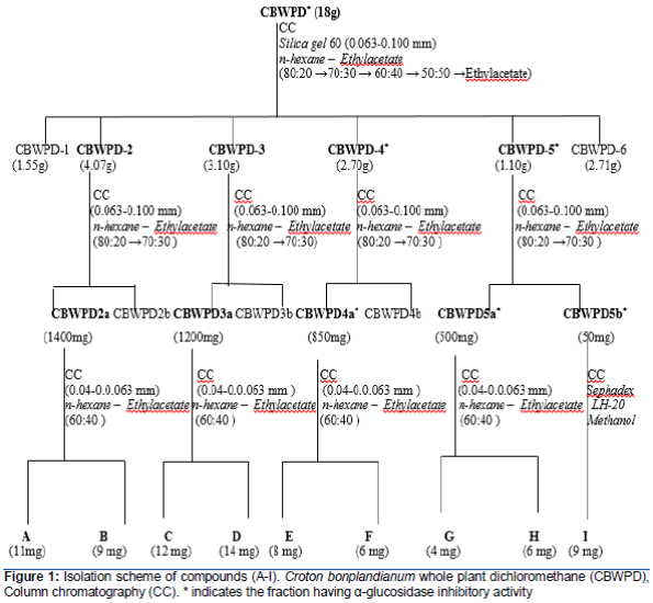
Dichloromethane extract (18 g) was subjected to column chromatography on silica gel using stepwise elution with n-hexane-ethyl acetate ((80:20 →70:30 → 60:40 → 50:50 →ethylacetate) in increasing order of polarity. six fractions (CBWPD 1- CBWPD 6) were obtained. The fraction CBWPD 2 (4.07g) subjected to column chromatography on silica gel using n-hexane-ethylacetate (80:20 →70:30) as eluent resulted two fractions (2a and 2b). The fraction 2a (1400 mg) was subjected to column chromatography on silica gel using n-hexane-ethylacetate (60:40) as eluent which gave two pure compounds A (11 mg) and B (9 mg). The fraction CBWPD-3 (3.10g) was subjected to column chromatography on silica gel using n-hexane-ethylacetate (80:20 →70:30) as eluent resulted two fractions (3a and 3b). The fraction 3a (1200 mg) was subjected to column chromatography on silica gel using n-hexane-ethylacetate (60:40) as eluent which gave two pure compounds C (12 mg) and D (14 mg).The fraction CBWPD-4 (2.74g) obtained by n-hexane-ethyl acetate (80:20 →70:30) was subjected to column chromatography on silica gel using n-hexane – EtOAc (60:40) as eluent resulted two fractions (4a and 4b). The fraction 4a (850 mg) was subjected to column chromatography on silica gel using n-hexane – EtOAc (60:40) as eluent which gave two pure compounds E (8 mg) and F (6 mg). The fraction CBWPD-5 (1.0g) was subjected to column chromatography on silica gel using ethylacetate-methanol (80:20 →70:30) as eluent resulted two fractions (5a and 5b). The fraction 5a (500 mg) was subjected to column chromatography on silica gel hexane – EtOAc (60:40) as eluent which gave two pure compounds G (7 mg) and H (6 mg). Fraction 5b (50 mg) was subjected to column chromatography on Sephadex LH-20 using methanol as eluent afforded compound I (9 mg). The structures of these isolated compounds were established by spectroscopic technique such as UV and IR spectroscopy. Proton Nuclear Magnetic Resonance (1H NMR), 13C NMR and Mass spectrophotometry (EIMS, HRMS) were used for elucidation of structure. Isolation scheme of compounds (A-I) from dichloromethane extract of whole plant of Croton bonplandianum (CBWPD) is given in .
In vitro α-glucosidase inhibition assay
The α-glucosidase inhibitory activity was assessed by a standard method [6], with slight modifications. A volume of 60 μl of sample solution and 50 μL of 0.1 M phosphate buffer (pH 6.8) containing α-glucosidase solution (0.2 U/mL) was incubated in 96 well plates at 37 ºC for 20 min. After pre-incubation, 50 μL of 5 mM p-nitrophenyl-α-D-glucopyranoside (PNPG) solution in 0.1 M phosphate buffer (pH 6.8) was added to each well and incubated at 37 ºC for another 20 min. The reaction was stopped by adding 160 μl of 0.2 M NaCO3 to each well, and absorbance readings were recorded at 405 nm by micro-plate reader and compared to control which had 60 μL of buffer solution in place of the extract. For blank incubation (to allow for absorbance produced by the extract), enzyme solution was replaced by buffer solution and absorbance recorded.
The concentrations of test compounds which inhibited the hydrolysis of substrates by 50 % (IC50) were determined by monitoring the effect of decreasing concentrations of these extracts in the assays on the inhibition values. IC50 values were then calculated using EZ-Fit Enzyme Kinetics program (Perrella Scientific Inc., Amherst, USA).
Statistical analysis
All data were analyzed by analysis of variance (ANOVA) and mean values were compared by Duncan’s Multiple Range Tests using SPSS software, version 15 (SPSS Inc, Chicago, IL, USA). P < 0.05 was considered statistically significant.
Results
Isolation of compound (A-I)
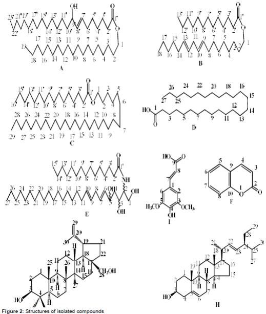
Dichloromethane extract was subjected to a series of column and flash chromatographic techniques as described in the experimental to obtain nine compounds reported for the first time from this species. These are identified as of n-pentacosanyl-n-nonadeca-7′-en-9′-α-ol-1′-oate (A), n-tridecanyl n-octadec-9,12-dienoate (B), nonacosyl hexadecanoate (C), heptacosanoic acid (D), 1,3,5-trihydroxy-2-hexadecanoylamino-(6e,9e)-heptacosdiene (E), cumarin (F), betulin (G), stigmasterol (H), and 3,5-dimethoxy 4-hydroxy cinnamic acid (I) respectively, on the basis of their spectral data, structure of isolated compound are given in .
n-Pentacosanyl-n-nonadeca-7′-en-9′-α-ol-1′-oate (A)
White amphorous powder (11 mg), m.p.158–159 °C; UV λmax MeOHnm (log ε): 224 (5.6) nm; IR (KBr) nmax cm-1:3487, 1751, 1618, 752 and 715 cm-1; 1H-NMR (CDCl3, 400 MHz) δ:; 5.31 (1 H, dd, J = 18.6, 5.2 Hz), 5.28 (1 H, brs), 4.21 (1 H, J = 11.1 Hz), 4.17 (1 H, J = 11.1 Hz) 3.87 (1 H, br’m), 2.75 (2 H, brs), 2.32 (1 H, d, J = 6 Hz), 1.65 (2 H, br’s), 1.23 – 1.28 (68 H, br’s), 0.88, 0.91 (6 H, brs, 2 CH3); 13C-NMR (100 MHz CDCl3) δ; 167.7, 130.2, 128.1, 75.8, 68.2, 62.1, 38.6, 32.1, 31.9 – 29.9, 24.7, 23.7, 23.1, 22.7, 14.2 and 14.1; EI-MS; 662 (18.2);HR-EI-MS m/z:662.3329 C44H86O3 (calculated. for C44H86O3; 662.3356);The physical and spectral data showed complete agreement with those reported in the literature [7].
n-Tridecanyl n-octadec-9,12-dienoate (B)
Amorphous solid (12 mg); m.p. 91-92 °C; IR; 1751 and 1648 cm-1 ;1H-NMR (CDCl3, 400 MHz)δ: 0.83 and 0.90 (t, each, 6H, J = 7.1 Hz), 1.23 – 1.27 (38 H, br’s), 2.42 (1H, d), d 2.23 (1 H, d), 4.39 (2 H, triplet, J =7.5 Hz), δ 4.1 (2 H, triplet, J = 7.0 Hz), d 5.23 (1H, dd, J = 14.9, 7.8 Hz); d 5.21 (1H, dt, J = 14.9, 7.8 Hz); d 5.18 (1H, dt, J = 15.1, 7.1 Hz); 5.05 (1H, dt, J = 15.1, 7.1 Hz); 13C-NMR (100 MHz CDCl3) δ:171.2, 135.6, 130.8, 128.9, 126.9, 68.1, 49.6, 38.7, 33.6, 31.9, 30.3 – 29.3, 28.6, 26.9, 24.1, 23.8, 22.9, 22.6, 10.9 and 18.5; EI-MS; 462 (12), 460 (14), 421 (27), 407 (19), 394 (13), 365 (15), 337 (11), 316 (29), 295 (20), 297 (45), 253 (17), 197 (27), 167 (79), 149 (89), 124 (100), 113 (55), 97 (51), 85 (65), 71 (83), 57 (98) and 43 (80); HR-EI-MSm/z:462.6685 C31H58O2 (calculated. For C31H58O2, 462.6689). The physical and spectral data corresponded to the reported values [8].
Nonacosylhexadecanoate (C)
Colorless amorphous solid (11 mg); m.p. 105-106 °C; IR;1652, 1615 and 1538 cm-1; 1H-NMR(CDCl3, 400 MHz) δ:0.87, 0.94 (6H, triplet, J = 6.8 Hz), d 1.25 – 1.38 (76H, br’s), 1.69 (quintet),d 2.03 (triplet, J = 7.2 Hz), d 4.21 (1H, d, J = 8.7 Hz); 13C-NMR (100 MHz CDCl3) δ:169.3, 69.1, 40.1, 33.0, 31.6 – 30.1, 24.9, 24.0, 11.4 and 14.4; EI-MS; 662 (25), 647 (55), 592 (21), 536 (18), 478 (23), 424 (19), 328 (21), 316 (15), 280 (20), 197 (21), 191 (17), 167 (26), 149 (78), 111 (19), 98 (33), 84 (43), 82 (41), 74 (65), 60 (78), 44 (95) and 42 (100); HR-EI-MSm/z:662.3479 (calculated. for C45H90O2; 662.3477). The physical and spectral data showed complete resemblance with the reported in the literature [9].
Heptacosanoic acid (D)
Amorphous solid; m.p. 86-87 °C; IR (KBr) nmax cm-1:3322, 2688 and 1721; 1H-NMR (CDCl3, 400 MHz) δ:0.91 (3H, triplet, J = 6.5 Hz), 1.59-1.61 (48H, br’s), 1.98 (2 H, quintet), 2.11 (2H, triplet, J= 7.3 Hz);13C-NMR (100 MHz CDCl3) δ : 176.1 (C-1), 34.9 (C-2), 32.1 (C-3), 29.6-29.9 (C-4-24), 25.6 (C-25), 22.9 (C-26), 14.2 (C-27); EI-MS m/z (rel. int.): 410 (21), 367 (23), 341 (35), 320 (19), 306 (13), 273 (17), 253 (18), 239 (17), 205 (19), 192 (22), 169 (32), 149 (44), 137 (54) 111 (61), 97 (78), 81 (82), 69 (100), 57 (88) and 41 (78); HR-EI-MSm/z:410.3782 (calcd. for C27H54O2, 410.3765). The physical and spectral data corresponded to the reported values [10].
Trihydroxy-2-hexadecanoylamino-(6E,9E)-heptacosdiene (E)
Gummy solid (16 mg); [a]D25: – 26.2° (c 0.10, pyridine); IR (KBr):3340, 3220, 1660, 1620 and 1540 cm-1 ; 1H-NMR (CDCl3, 400 MHz) δ: 0.89, 0.94 (6H, triplet, J = 6.8 Hz), 1.28 (18H, br s), 1.32 (18H, br s), 2.03 (2H, t, J = 7.0 Hz, H-4), 2.15 (2H, t, J = 7.0 Hz, H-2’), 3.41 (2H, m, H-8), 3.59 (1H, m, H-3), 3.65 (1H, dd, J= 11.5, 5.0 Hz, H-1b), 3.94 (1H, m, H-5), 4.22 (1H, dd, J= 11.3, 4.9 Hz, H-1a), 4.87 (1H, dd, J = 15.5, 8.4 Hz); d 4.91 (1H, dt, J = 15.5, 8.4 Hz); d 5.05 (1H, dt, J = 16.1, 6.9 Hz); 5.18 (1H, dt, J = 16.1, 6.9 Hz); 13C-NMR (100 MHz CDCl3) δ:169.4, 133.6, 132.4, 130.7, 129.8, 82.4 (C-3), 74.7 (C-5), 69.1 (C-1), 57.0 (C-2), 33.1 (C-4), 30.4 (C-8), 31.6, 30.8, 30.4, 24.9, 24.0, 23.7, 21.6, 14.4, 11.4; EI-MS:m/z (rel. int. %) 663 (12), 438 (32), 423 (28), 379 (42), 335 (39), 305 (21), 292 (33), 279 (19), 225 (62); HR-FAB-MS (+ive): m/z 664.6246 (calculated. for C42H82NO4; 664.6249). The physical and spectral data showed complete agreement to the reported values [11].
2 H-1-Benzopyran-2-one (F)
Colourless crystalline solid (11 mg); m.p. 70 oC; UV λmax MeOHnm (log ε): 298 (4.01), 238 (3.31), 225 (2.99); IR (KBr) nmax cm-1: 1725, 1623, 1589, 1512; 1H-NMR (CDCl3, 400 MHz) δ:6.41 (1H, d, J= 9.5 Hz, H-3), 7.25 (1H, d, J= 8.7 Hz, H-5), 7.49 (1H, d, J= 8.7 Hz, H-8), 7.52 (2H, m, H-6, H-7), 7.68 (1H, d, J= 9.5 Hz, H-4); EI-MS m/z (rel. int.): 146.0 (29); HR-EI-MS m/z:146.0534 (calcd. for C9H6O2,146.0538).The physical and spectral data corresponded to the reported values [12].
Betulin (G)
Crystallized from MeOH (20 mg); m.p. 251-252°C; [α]D25: + 20.4° (c 0.42, C5H5N); IR (KBr) nmax: 3435, 3070, 1635, 880 cm-1; 1H-NMR (400 MHz; CDCl3) δ: 4.68 (2H, m, H-29), 3.75 (1H, dd, J = 10.7, 4.2 Hz, H-3), 3.81, 3.42 (1H each, d, J = 11.0 Hz, H-28), 1.68 (3H, br. s, CH3-30), 1.02 (3H, s, CH3-26), 0.98 (3H, s, CH3-27), 0.92 (3H, s, CH3-24), 0.89 (3H, s, CH3-23), 0.87 (3H, s, CH3-25); EI-MS (rel. int. %) m/z: 442 [M]+ (15); HR-EI-MS m/z: 442.3814 (calculated for C30H50O2; 442.3818);The physical and spectral data corresponded to the reported values [13].
Stigmasterol (H)
Colorless crystalline solid (17 mg); m.p. 170-171 °C; [α]D25; -51.5˚ (c = 0.28, CHCl3); IR (CHCl3) nmax cm-1: 3432 (OH), 1648 (C = C); 1H-NMR (CDCl3, 400 MHz) δ: 5.33 (1H, m, H-6), 5.15 (1H, dd, J = 15.2, 8.4 Hz, H-22), 5.02 (1H, dd, J = 15.2, 8.6 Hz, H-23), 3.28 (1H, m, H-3), 0.90 (3H, d, J = 6.5 Hz, Me-21), 0.83 (3H, d, J = 6.6 Hz, Me-26), 0.84 (3H, t, J = 7.0 Hz, Me-29), 0.81 (3H, d, J = 6.5 Hz, Me-27), 0.80 (3H, s, Me-19), 0.65 (3H, s, Me-18); EIMS m/z (rel. int. %): [M]+ 412 (8) HREIMS m/z: 412.3919 (calculated for C29H48O, 412.3930).The physical and spectral data showed complete resemblance with the those reported in the literature [14-15].
3,5-dimethoxy 4-hydroxy cinamic acid (I)
Yellow solid from acetone (15 mg); m.p. 89-90 0C; UV λmax MeOH nm (log ε): 329 (4.01), 239 (3.92), 205 (3.85) nm; IR (KBr)υmax cm-1: 3363, 1704, 1663, 1604, 1449 cm−1; 1H-NMR (CDCl3, 400 MHz):7.58 (1H, doublet, J =16 Hz), 6.88 ( 2 H, singlet), 6.32 (1 H, doublet, J = 16 Hz) , 4.85 (OH ) and 3.86 (6 H , singlet); 13C-NMR (CDCI3, 100 MHz): 170.87, 149.47, 147.11, 139.51, 126.74, 116.37, 106.85, 147.11, 116.36, 106.84 and 56.85. EIMS m/z (rel. int) %: 224 (38), 196 (36), 190 (45), 161 (34), 149 (45), 131 (12), 119 (24), 107 (15) and 78 (49); HREIMS m/z M+ 224.1233(calculated for C11H12O5; 224.1241). The physical and spectral data corresponded to the reported values [16].
In vitro α-glucosidase inhibition
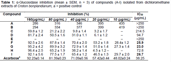
All the isolated compounds were screened for α-glucosidase inhibitory activity. Compound 2H-1-Benzopyran-2-one (F), Betulin (G) & 3, 5-Dimethoxy 4-hydroxy cinnamic acid (I) possessed significant α-glucosidase inhibitory activity in a concentration-dependent manner, and showed more potent inhibitory activity, with IC50 values ranging from 23.0 to 26.7 μM, than that of a positive control acarbose (IC50, 38.2 μM). Results are summarized in .
Discussion
Diabetes is one of the world's greatest health problems, affecting about 171 million people and most of these will be dominated by those suffering from type II diabetes [17]. One of the strategies to monitor blood glucose for type II diabetes mellitus is to either inhibit or reduce the production of glucose from the small intestine. Diet rich in carbohydrate causes sharp rise in the blood glucose level as the complex carbohydrates in the food is rapidly absorbed in the intestine aided by the α-glucosidase enzyme which breaks disaccharides into absorbable monosaccharides, α-glucosidase inhibitor inhibits the disaccharide digestion and impedes the postprandial glucose excursion to enable overall smooth glucose profile [18]. Thus, natural products of great structural diversity are still a good source for searching for such inhibitors, thereby motivating to explore biologically active compounds from the highly diverse plants.
The development of the alpha-glucosidase inhibitor provides a new approach in the management of diabetes. By competitive and reversible inhibition of intestinal alpha-glucosidases, alpha-glucosidase delays carbohydrate digestion, prolongs the overall carbohydrate digestion time, and thus reduces the rate of glucose absorption. After oral administration of alpha-glucosidase, the postprandial rise in blood glucose is dose-dependently decreased, and glucose-induced insulin secretion is attenuated. Due to diminished postprandial hyperglycemia and hyper-insulinemia by alpha-glucosidase inhibitor, triglyceride uptake into adipose tissue, hepatic lipogenesis, and triglyceride content are reduced. Therefore, alpha-glucosidase inhibitor treatment not only flattens postprandial glycemia, due to the primary and secondary pharmacodynamic effects, but also ameliorates the metabolic state in general.
Thus, alpha-glucosidase inhibitor may have the potential to delay or possibly prevent the development of diabetic complications [19]. Compound 2H-1-Benzopyran-2-one (F), Betulin (G) & 3, 5-Dimethoxy 4-hydroxy cinnamic acid (I) possessed significant α-glucosidase inhibitory activity in a concentration-dependent manner, and showed more potent inhibitory activity, with IC50 values ranging from 23.0 to 26.7 μM, than that of a positive control acarbose (IC50, 38.2 μM). It has been displayed that these compounds are attractive candidates for α-glucosidase inhibitory activity.
Conclusion
In the light of these findings, the isolated compounds exhibited inhibitory activity against α-glucosidase in a dose-dependent manner and therefore may potentially be developed as new α-glucosidase inhibitors for the treatment of type 2 diabetes.
Declarations
Acknowledgement
References
Archives


News Updates